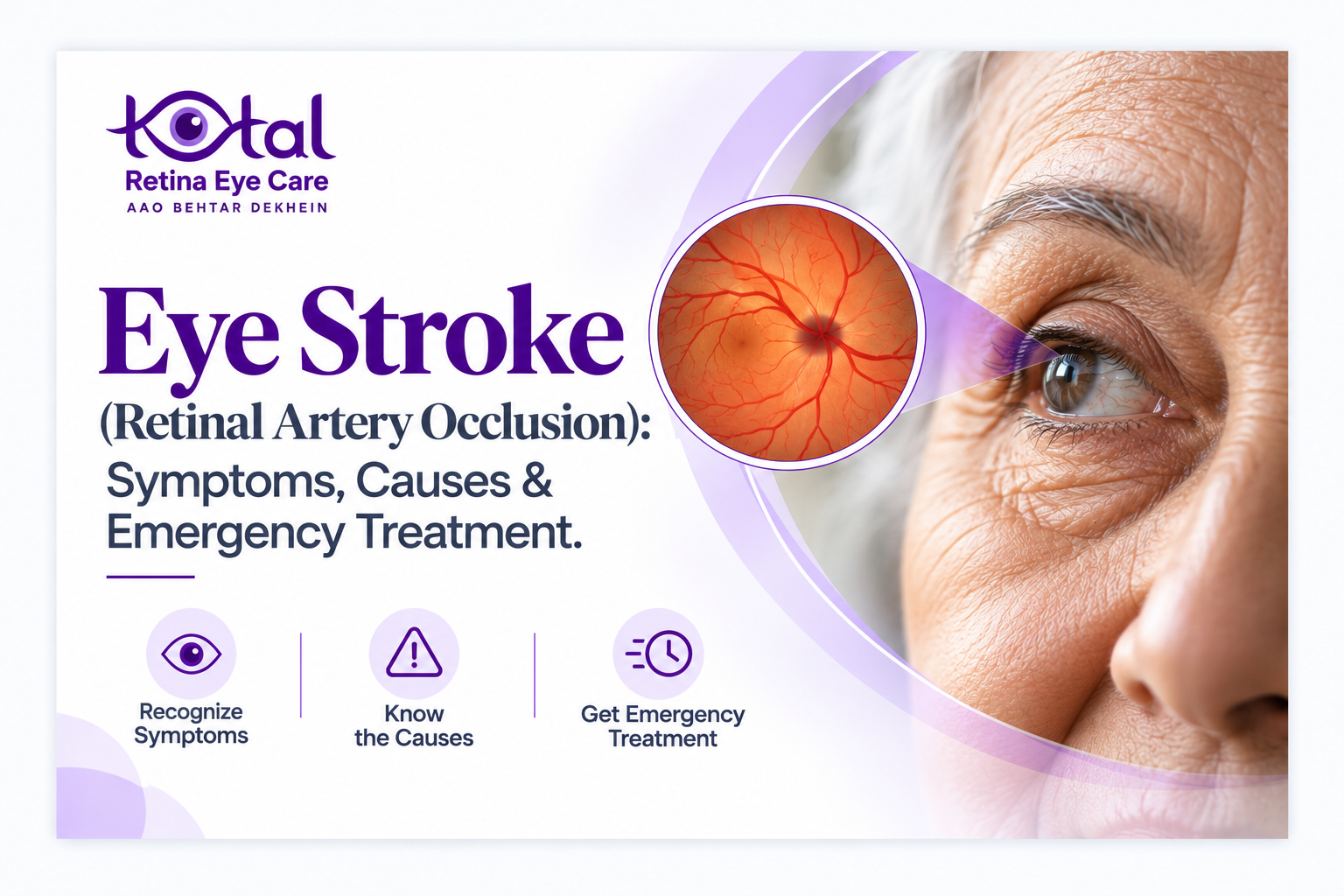
Picture this. You wake up, cover one eye while brushing your teeth, and realize the other one can barely see. There is no pain, no redness, nothing that looks wrong. Many people shrug it off and decide to “wait and see.” With an eye stroke, that wait can cost you your sight.
An eye stroke, known medically as retinal artery occlusion (RAO), happens when the blood supply to the retina is suddenly blocked. It is one of the true emergencies in eye care, and it is also a warning that something may be wrong with your heart or blood vessels.
In this guide, our retina team at Total Retina Eye Care, an eye hospital in Nagpur, explains what an eye stroke is, what it feels like, why it happens, how it is treated, and what you can do to lower your risk.
If you have sudden vision loss in one or both eyes, go to the nearest hospital emergency department immediately. Do not wait for an appointment.
What Is an Eye Stroke?
The retina is a thin layer of light-sensing tissue at the back of the eye. It works like the film in a camera, turning light into signals that the brain reads as images. It is also one of the most oxygen-hungry tissues in the body.
The retina receives blood mainly through the central retinal artery and its branches. When a clot, a piece of cholesterol plaque or another particle (called an embolus) lodges in one of these arteries, blood flow stops. Retinal cells begin to lose function quickly, and the longer the blockage lasts, the more likely the damage becomes permanent.
The brain and the retina share a similar blood supply and are both part of the central nervous system. For this reason, doctors now treat a retinal artery occlusion much like a stroke of the brain.
Types of Retinal Artery Occlusion
Central Retinal Artery Occlusion (CRAO)
The main artery is blocked. Vision loss is usually severe and affects most of the eye. Many patients are left with only the ability to count fingers, see hand movements or perceive light.
Branch Retinal Artery Occlusion (BRAO)
A smaller branch is blocked, so only part of the retina is affected. People may notice a dark patch, a missing section of the visual field or a shadow in one area. Central vision is often (but not always) better preserved than in CRAO.
Transient Retinal Artery Occlusion (Amaurosis Fugax)
Sometimes the blockage clears on its own. Vision goes dim or dark in one eye, like a curtain falling, for a few seconds to several minutes, and then returns. It is tempting to ignore this because everything seems normal afterwards. Please don’t. It is a “mini-stroke” of the eye and can be a warning of a bigger event to come.
Symptoms of an Eye Stroke
The hallmark is sudden, painless loss of vision in one eye. Other signs include:
• A dark curtain or shadow across part or all of your vision
• Sudden blurring
• A blind spot or missing patch of vision
• Loss of side vision
• Brief episodes of vision going dark that clear within minutes
Because there is no pain, people often delay care. Remember: painless does not mean harmless.
What Causes a Retinal Artery Occlusion?
In most patients the cause is an embolus travelling from elsewhere in the body. The most common sources are:
• Carotid artery disease: plaque in the large neck arteries can break off and travel to the eye
• Heart conditions: irregular heartbeat such as atrial fibrillation, damaged heart valves, or clots forming in the heart chambers
• Atherosclerosis: hardening and narrowing of arteries from long-standing high blood pressure, diabetes and high cholesterol
Less commonly, an occlusion may be caused by:
• Giant cell arteritis (GCA): inflammation of blood vessels, mainly in people over 50. It needs urgent treatment with steroids to protect the other eye. Warning signs include new headache, jaw pain while chewing, scalp tenderness, fever and unexplained weight loss
• Blood clotting disorders
• Inflammatory or autoimmune diseases
• Certain medicines or drugs, and eye or facial trauma
• Rare conditions in younger people, such as heart defects
Who Is at Risk?
Retinal artery occlusion is most common in people in their 60s and 70s, but it can occur at any age. You are at higher risk if you have:
• High blood pressure
• Diabetes
• High cholesterol
• Heart disease or atrial fibrillation
• Carotid artery disease
• A previous stroke or TIA
• A history of smoking
• Obesity or a sedentary lifestyle
• A blood clotting disorder or an inflammatory disease
Eye Stroke and Brain Stroke: The Connection
This is the part many patients do not expect. An eye stroke is not just an eye problem. People who have had a retinal artery occlusion face a higher risk of stroke, heart attack and other vascular events. The risk is highest around the time of the occlusion, from about two weeks before it to roughly a month after. Some patients already have small silent strokes in the brain by the time they are seen.
This is why current guidelines (including the American Academy of Ophthalmology’s 2025 Preferred Practice Pattern) recommend that a patient with a sudden retinal artery occlusion be assessed urgently at an emergency department that can also manage stroke, with brain imaging, a check of the neck arteries, a heart rhythm check and blood tests. Treating the eye and protecting the brain go hand in hand.
How Is an Eye Stroke Diagnosed?
At our eye hospital in Nagpur, a retina specialist usually makes the diagnosis based on your story and a careful examination. The tests may include:
• Dilated retinal examination: the retina often looks pale and swollen, and in CRAO a “cherry-red spot” may appear at the centre of the macula
• Optical coherence tomography (OCT): a scan that shows swelling and thinning of the retinal layers
• Fundus photography: records the appearance of the retina
• Fluorescein angiography: shows delayed or absent blood flow in the retinal vessels
• Visual field testing: maps the areas of lost vision
Because the cause usually lies outside the eye, you will likely also need:
• Blood pressure and blood sugar checks
• Cholesterol and blood clotting tests
• ESR and CRP (especially if you are over 50, to look for giant cell arteritis)
• Carotid Doppler ultrasound or CT/MR angiography
• ECG and heart rhythm monitoring
• Echocardiography
• Brain imaging (MRI or CT)
Treatment: What Can Be Done?
We want to be honest here: there is currently no treatment proven to reliably restore vision after a central retinal artery occlusion. That is why speed matters so much and why prevention is so important.
The retina has very little tolerance for lost blood supply, so the first hours matter. Current guidance from the American Academy of Ophthalmology is that anyone with a sudden retinal artery occlusion should be sent immediately to an emergency department linked to a stroke center for assessment, and that after the acute phase there are no proven treatments to reverse the vision loss. In practice, care focuses on three things:
• A rapid stroke-style evaluation: brain and vessel imaging, heart rhythm monitoring and blood tests to find the source of the blockage and look for silent strokes
• Treatment for giant cell arteritis when suspected: in patients over 50, high-dose steroids are started promptly, without waiting for a biopsy, to protect the vision in the other eye
• Secondary stroke prevention: antiplatelet medicines or blood thinners where appropriate, statins, blood pressure and diabetes control, and treatment of carotid or heart disease
What about clot-busting drugs? For some years, intravenous thrombolysis (such as alteplase) given within 4.5 hours was considered a possible option, and the 2021 American Heart Association statement reflected that. Since then, two randomised trials have changed the picture. The THEIA trial (alteplase versus aspirin) and the TenCRAOS trial (tenecteplase versus aspirin) both found no meaningful improvement in vision with thrombolysis, and TenCRAOS reported more adverse events, including a brain bleed in one patient. Because of this, routine thrombolysis is no longer widely supported for CRAO. Whether it should ever be used in carefully selected patients is being decided by stroke teams as further trial data (such as the REVISION trial and combined analyses) are reviewed.
What about eye massage, eye-pressure medicines and similar measures? These have long been used in emergency rooms, but there is no strong evidence that they restore vision. They should never replace, or delay, transfer to hospital.
Please do not try home remedies, and do not delay hospital care to look for one. Time is the single most important factor, because it determines which options are open to you and how quickly the cause can be found and treated.
Can Vision Recover?
It depends on the type and severity of the blockage and how quickly blood flow returns. In CRAO, many patients are left with significant permanent vision loss in the affected eye, though a minority recover some useful sight. In BRAO, outcomes are often better because only part of the retina is affected.
Even when the vision does not return, follow-up care is worthwhile. We help patients make the most of the vision they have, protect the other eye, and bring their vascular risk factors under control. Some people also develop abnormal new blood vessels later on, which we can monitor and treat.
How to Reduce Your Risk
Most of the risk factors for eye stroke are the same as for heart attack and brain stroke, and most can be managed:
1. Control your blood pressure. Check it regularly and take medicines as prescribed.
2. Manage diabetes. Keep your blood sugar within the targets set by your doctor.
3. Lower cholesterol through diet, exercise and medicines if needed.
4. Stop smoking. This is one of the most effective steps you can take.
5. Stay active and keep a healthy weight.
6. Treat heart rhythm problems such as atrial fibrillation.
7. Do not skip check-ups. If you have diabetes, high blood pressure or heart disease, have a dilated retinal exam at least once a year, or as often as your doctor advises.
8. Act on warning episodes. Any temporary loss of vision, even if it clears, needs same-day medical evaluation.
When to Go to the Emergency Room
Seek emergency care immediately if you experience:
• Sudden loss of vision in one eye, partial or complete
• A dark curtain or shadow moving across your vision
• Brief episodes of vision loss that go away on their own
• Sudden vision loss along with weakness, facial drooping, speech difficulty or severe headache. Call for an ambulance right away
When you go, tell the staff clearly: “I have sudden loss of vision in one eye.” Mention the exact time you first noticed it. This helps the team decide quickly which treatments are possible.
Frequently Asked Questions
1. Is an eye stroke painful?
No. Typically it causes sudden, painless loss of vision. The lack of pain is the reason many people delay seeking help.
2. How is an eye stroke different from a brain stroke?
Both are caused by blocked blood flow. In a brain stroke, the blockage is in an artery supplying the brain. In an eye stroke, it is in an artery supplying the retina. Having one raises the risk of the other, which is why a full vascular check-up is needed.
3. How long do I have to get treatment?
As little as possible. Go to the emergency department as soon as you notice the vision loss, and note the time it started. Even if many hours have passed, you still need urgent evaluation to find the cause and prevent a brain stroke.
4. Can an eye stroke happen in young people?
Yes, although it is less common. In younger patients, doctors look for heart defects, clotting disorders, inflammatory conditions, migraine-related causes, trauma or drug use.
5. Can an eye stroke happen again?
It can, in the same eye or the other eye, which is why controlling the underlying cause is so important.
6. Is an eye stroke the same as a retinal detachment?
No. A retinal detachment is when the retina peels away from the back of the eye, often with flashes, floaters and a shadow. A retinal artery occlusion is a blood supply problem. Both are emergencies that need immediate evaluation by an eye specialist.
7. Will my vision come back on its own?
Sometimes the blockage clears in minutes (transient episodes), but in a true occlusion the loss often persists. You should never assume it will resolve. Get checked straight away.
8. Final Thoughts
An eye stroke is silent, sudden and easy to underestimate. But it is also a clear signal from your body that your blood vessels need attention. If you or a family member notices sudden vision loss, treat it exactly as you would a stroke: go to the emergency department without delay.
At Total Retina Eye Care, an eye hospital in Nagpur, our vitreoretinal specialists work alongside emergency, neurology and cardiology teams to evaluate retinal vascular emergencies and to help patients protect their vision and their long-term health. If you have diabetes, high blood pressure or heart disease, a regular retinal examination is a simple step that can make a real difference.
If you have sudden vision loss, do not wait for an appointment. Go to the nearest emergency department now. For follow-up retinal care after an emergency, or for regular retinal check-ups if you have diabetes, high blood pressure or heart disease, you can consult a retina specialist in Nagpur at Total Retina Eye Care.